

Long-term brain observations are now possible due to a new device created by University of Minnesota researchers.
Research released on April 2 by the University’s mechanical engineering department introduced a new 3D-printed skull implant to help study the brain for longer durations. Researchers said they can observe anything from Alzheimer’s and Parkinson’s disease to concussions and drug use.
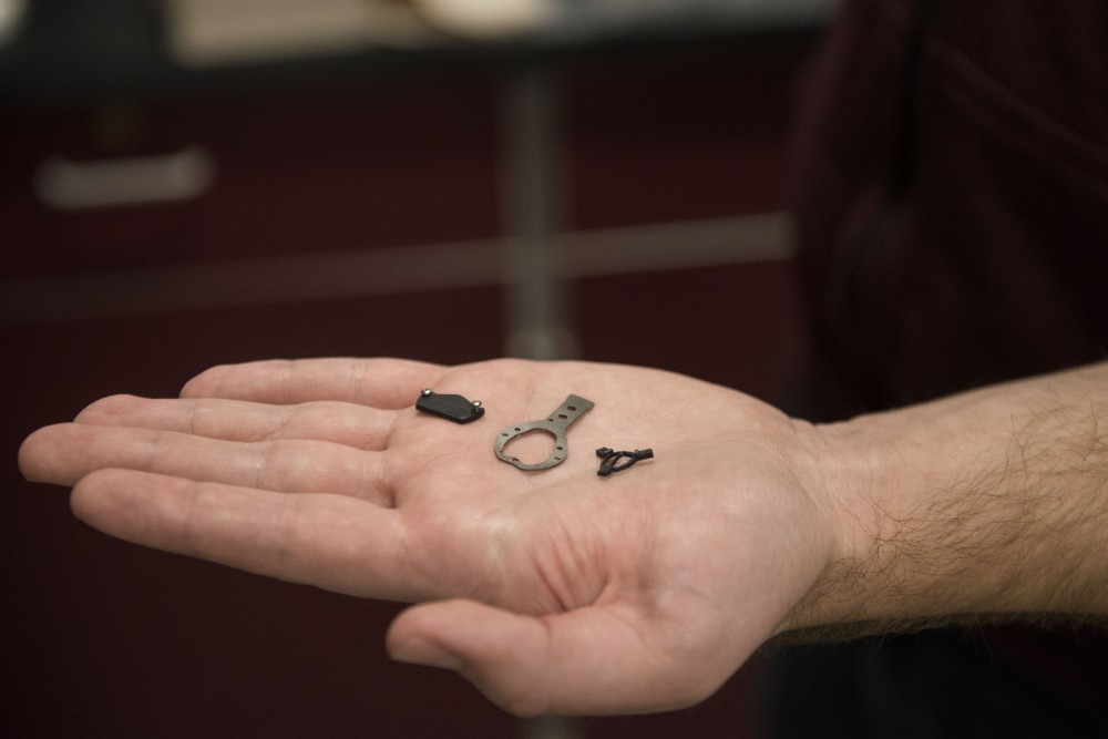
The See-Shell device is a thin plastic film bonded to a 3D-printed frame, which contours to the shape of the skull and acts as a “window” into the brain. So far, the device has only been used on mice, which have similar brain structures to humans.
“The windows allow us to do a type of interrogation of the brain … that we’ve just not been able to do before,” said Timothy Ebner, head of the University’s Department of Neuroscience. “We can image the activity across the brain; we can go down and look at the individual cells.”
The team observed mice’s brain activity for over 10 months. The adaptability of the 3D design sets this device apart from previous attempts to observe the brain, researchers said.
Suhasa Kodandaramaiah, senior author of the study and assistant professor of mechanical engineering, said, “we really needed a way to sort of profile the skull and replace that with something that exactly matches this sort of contour.”
Researchers are able to potentially find each neuron roughly 500 times per second, much faster than an MRI, Kodandaramaiah said. He said the optical imaging allows them to zoom in on features as small as a micrometer.
Their current research is looking into how different parts of the brain interact depending on different behaviors.

“Neuroscientists have been facing this critical challenge of monitoring large regions of the brain at the same time at high resolution,” said lead author Leila Ghanbari, fourth-year Ph.D. candidate in mechanical engineering. “We thought about devices that give us that optical access … to large brain regions at the same time … how they collaborate with each other, how they perform their functions. And we also wanted to see the structure of the brain.”
The researchers are currently using the device to analyze how repeated concussion injuries can impact multiple parts of the brain. Kodandaramaiah said injuries happening in one part of the brain can also be seen globally.
“We have been able to show how they’re different parts of the brain that we’re imaging in the mouse, how there is a network of areas and how they are all working together when mice, for example, engage in different behaviors,” Ebner said. “It’s something very analogous to what we see in humans.”
Clarification: Researchers are able to potentially find each neuron roughly 500 times per second.
Clarification: Suhasa Kodandaramaiah is the senior author of the study.