

For University of Minnesota Biomedical Engineering Professor Bin He , when it comes to studying the brain for epilepsy, 10 seconds is not fast enough. ThatâÄôs how long it takes to get a reading from a functional magnetic resonance imaging (fMRI ) system, but with neurons firing at one millisecond, Professor HeâÄôs research seeks to develop a new way that would offer a faster, more precise way to look at the brain. HeâÄôs research looks to integrate two widely known brain scanning techniques, Electroencephalography (EEG) EEG and fMRI, into one method.

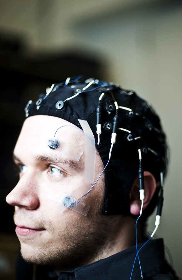
With an integration of fMRI and EEG, He believes a detailed mapping of the brainâÄôs connectivity is possible. Functional neuroimaging allows researchers and doctors to see the brainâÄôs processes by detecting activity. Areas of activity are shown in color on the scan, pinpointing exactly where the process took place. With more precise readings, doctors may be able to better treat patients suffering from neurological and mental disorders. According to He, fMRI and EEG are currently two predominant methods of mapping brain activity. With EEG, electrode sensors are placed on the scalp in order to monitor brain activity. In fMRIâÄôs, a patient will lie in an MRI machine while their head is scanned. These two methods are the counterparts of each other. EEG gives better precision of a measurement with respect to time in the reading but is less exact in location; fMRI is the opposite. One important aspect of this research is that it is non-invasive; meaning researchers and doctors would not need to open a patientâÄôs skull in order to perform the reading. He cited the use of non-invasive techniques as a personal philosophy for all of the research conducted in his lab. A researcher on epilepsy, He said the use of non-invasive techniques is a better answer to current methods of treating patients. When doctors attempt to take a scan of a personâÄôs brain who has epilepsy, He said their skull is opened and an electrode grid is placed on the brain. The person is then placed in the intensive care unit for one to two weeks until they have a seizure and doctors can determine the location of activity in the brain during the seizure. With HeâÄôs new techniques in functional neuroimaging, a person could wear a cap covered in electrode sensors which would record activity during a seizure. He said he believes the person would not even need to stay in the hospital âÄî they could take the cap home. He said that currently, readings on epilepsy patients do not necessarily catch all of the areas that are active when a seizure occurs. He said many times doctors will remove the part of the brain determined to be âÄúbadâÄù in epilepsy patients, only for the patient to experience more seizures months later due to another area of the brain, which was missed, being affected by the disorder. âÄúPatients are constantly living in fear,âÄù Christopher Wilke, a fourth-year biomedical engineering MD/PhD student who works in HeâÄôs lab, said. Using HeâÄôs technique in epilepsy detection would allow doctors and patients to make better educated decisions through more exact data, Wilke said. âÄúBefore, people thought epilepsy comes from a bad spot. They cut it out, but a few months later another seizure, another bad spot,âÄù He said. âÄúIt is not just a spot, it is a network. If you can map that network you can treat it.âÄù It is the lack of knowledge in the area of brain connectivity that He said allows for doctors to miss other bad spots when treating patients. With the University being a pioneer in connectivity research, He said understanding how the different regions of the brain communicate with each other is at the center of his research. A man of many interests, HeâÄôs lab at the University currently has 20 people of various levels of experience working in it who are divided between four different research efforts, functioning neuroimaging being one of them. Heavily funded by various groups like the National Institute of Health, the National Science Foundation, the state of Minnesota, the University and a partnership with Mayo Clinic, He said his labâÄôs research costs more than $1 million a year. His research on neuroimaging has also created working collaborations with professors from other departments on campus, such as psychology professor Stephen Engel, moving beyond the realms of biomedical engineering and mental disease Engel, who researches how learning can change the connections in the brain, said HeâÄôs work provides him with stronger results. Engel added that when choosing someone to collaborate with, He stood out because Engel needed the best technique to measure the brain, which He could provide him. âÄúIâÄôm really grateful to him,âÄù Engel said. The symbiotic collaborations also allow He to test the new technique, which he would like to see made available to the entire scientific and medical community.