A team headed by researchers at the University of Minnesota has developed a new kind of Magnetic Resonance Imaging (MRI) machine that produces images similar in quality to a standard machine while taking up much less space.
This new machine separates into four pieces and fits into the bed of a truck, allowing it to be driven to remote communities where residents may not ordinarily have access to MRI scanners. A standard MRI machine weighs 4,500 kilograms; the new one only weighs 400.
Michael Garwood, associate director of the Center for Magnetic Resonance Research (CMRR) and lead researcher on the project, said 90% of the world’s population does not have access to an MRI machine.
Dependable access to an MRI machine is important for helping diagnose, monitor and treat injuries and diseases, according to Garwood.
“This is the motivating factor for all this work, there’s just not enough MRIs,” Garwood said. “You can’t access 90% of the world’s population with it and it’s time to do something about that.”
Unlike a standard machine, Garwood’s MRI only scans the head, which is one of the reasons this new machine can be as small as it is and still function properly.
The machine looks like a hooded hair dryer from a salon. The patient sits in the chair, which is raised until their head is fully under the dome. There is a strip of glass placed at eye level for the patient to look out of during the procedure, reducing feelings of claustrophobia.
Garwood said keeping children in mind when designing the machine was a priority. Kids can get scared when going through the larger machine, so Garwood wanted to make the MRI process more inviting and less intimidating.
Garwood said he originally designed the machine to run at 1.5 tesla, a unit of measurement for magnetic flux, just like a standard MRI machine. However, by using paraffin wax as a binding agent instead of epoxy, the machine was able to run at about half power.
Even though it is a lower operating field than standard MRI machines, Garwood said, his machine will not affect the images it generates and is safe for human use.
On a personal tour of the lab, Garwood showed off the machine’s inner workings. When asked for a photo, he opted to raise himself into the machine while the magnet was on, performing what was essentially the first unofficial human trial.
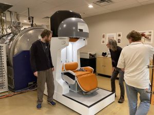
Despite Garwood not taking a scan of his brain while having his photo taken, it was a demonstration of how close the team is to beginning actual testing on people. Controlled human trials, Garwood estimated, will begin sometime in the next two months.
Initially, the researchers had only performed scans on water bottles and lemons in the machine. These scans still yielded high-quality images with no need to upscale them with artificial intelligence (AI).
The project required collaboration from researchers within the University, Yale, Columbia and out-of-country scientists from Brazil and New Zealand.
One of Garwood’s professor colleagues from Brazil, Alberto Tannus, designed a spectrometer that acts as the brain of the machine. It was designed separately from Garwood’s project and was originally meant to act as a research instrument.
Contained in a 1-inch-by-1-inch processing chip, the spectrometer creates interconnections between logical ports. Like calling a general customer service line and being re-routed to a more specific department, the spectrometer helps the machine get information where it needs to go.
Tannus said the research was hard to conduct because of funding. In Brazil specifically, Tannus said he was dealing with a shy budget.
“Something that I learned is if you can do it with this very low budget here in Brazil, you can do it with any amount of money anywhere in the world,” Tannus said.
Ben Parkinson, a researcher on Garwood’s team from New Zealand, said when the team had to design a magnet for the machine, they had to use a different process because of how small their machine is compared to a traditional MRI.
Garwood’s machine can produce clinical-quality images at a smaller size because it only gathers images from the head. Its smaller size and lower tesla also allow it to have a less uniform magnetic field than a standard MRI machine.
Parkinson said another difference between their machine and a standard MRI is the temperature control. A standard machine needs the coils inside the magnet to be cooled off in a bath of liquid helium.
Garwood’s MRI uses a high-temperature superconducting (HTS) magnet, which helps the coils inside cool faster and more efficiently.
Cristoph Juchem, an associate professor at Columbia University in New York, designed the hardware for the coils inside of the magnet.
A traditional MRI machine uses gradient coils to predictably manipulate the magnetic fields and typically relies on a set of three coils. The array Juchem developed houses 31 coils that can be controlled individually to create more flexible and dynamic patterns.
Juchem said getting a project like this off the ground is a hard enough task and is very excited they are nearing human trials.
“The fact that we get clean images, even though it’s not human yet, means we’re certainly on the right track and now the next goal is to do this on people like you and me,” Juchem said.





Gerard
Dec 18, 2023 at 7:02 am
Thank goodness that somebody finally designing machines that people prefer and can easily use instead of those horrible tunnel MRI machines that most people hate by just looking at them !
I hope you can market these great miniature machines instead of the older MRI design.
I hope hospitals and other medical companies start buying these new machines soon.
It is about time that patient comfort and ease of use is more important than how many Teslas the machine has !
Claustrophobia is no joke !