
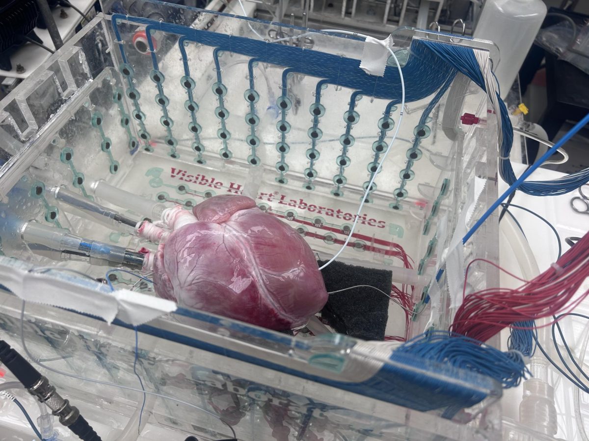
Tucked in the basement of the Mayo Building on the University of Minnesota’s Twin Cities campus sits a detached, beating pig’s heart floating in clear liquid.
Graduate and undergraduate students surround it, observing the heart directly or through screens displaying a live stream of its insides.
Visible Heart Laboratories (VHL) reanimates more than 200 hearts a year, mainly from animals, laboratory director Paul Iaizzo said. Occasionally the lab receives human hearts to work on, having reanimated 98 human hearts as of February.
The lab also works on other organs, like kidneys and lungs, Iaizzo said.
VHL began in 1997 when Iaizzo and his coworkers started studying mammalian hearts, or hearts belonging to mammals, according to the lab’s website. It is a collaboration between the University and Medtronic to research the normal functions of living organisms and their body parts.
The lab has a rich history, Iaizzo said. The first battery-powered pacemaker was invented in the same space in the late 1950s by University researcher Earl Bakken. The walls are adorned with photos of past researchers who used the space before VHL, alongside past and current students who have worked in the lab.
The heart sits inside a tub called the Visible Heart apparatus and is partially submerged in a clear liquid blood substitute called Krebs-Henseleit. The heart is hooked up to machines mimicking arteries and veins to control the amount of blood flowing into the heart, and a heater helps to keep the blood substitute at body temperature allowing the liquid to oxygenate.
The blood substitute lacks red blood cells, which means the heart can only beat for a few hours, Iaizzo said.
“The clear solution allows us to see what’s happening inside the heart,” Iaizzo said.
The researchers place cameras inside and around the heart to see the insides of a working heart to understand its anatomy, Iaizzo said.
Occasionally, the lab livestreams its research to viewers worldwide so others can benefit from their findings, Iaizzo said.
The pig heart is much bigger than a human heart but is structurally similar to a human one, Iaizzo said. This means research conducted on a pig heart can translate to healthcare advancements for human medicine.
Down the hall, VHL houses a library of preserved hearts dating back to 2000. The hearts sit in jars on shelves lining the walls of the room.
Iaizzo said because VHL is part of an academic institution, anyone can come in and look at the hearts in the library to further their research.
The laboratory also develops free augmented reality tools for educational use so students can better understand the heart’s structure, Iaizzo said.
“The first time a student works on an actual body is very different from working on a cadaver,” Iaizzo said. “This gives students the chance to see what a living body looks like because that is visually more similar to what surgery looks like.”
VHL also makes 3D-printed replicas of the hearts in the library, Iaizzo said.
“Every heart is different, so being able to show students models of different hearts is beneficial,” Iaizzo said.
The 3D models can also be used to explain surgery to patients and their loved ones so they have a better understanding of the procedure, Iaizzo said.
“Being able to show families exactly what will happen is comforting to them,” Iaizzo said.
Amanda DeVos, a doctoral student studying biomedical engineering, said working in VHL gave her an advanced understanding of how the heart and other organs function.
“What I learn working on one organ can end up informing my work on another,” DeVos said.
DeVos said the experiences she gained at VHL are unique and difficult to replicate elsewhere.
“Every day is different and we’re always working on something new,” DeVos said.